Research Facilities
Baycrest has advanced neuroimaging and research technology available to Baycrest researchers.
These facilities are also available to other organizations, companies and universities for a fee to support their research and development work. For more information, email [email protected].
Motor vehicle accidents remain a leading cause of accidental death worldwide. Death and injury rates are particularly high for young inexperienced drivers and elderly drivers. Understanding the behavioral changes that are associated with maturation and aging could inform assessments of driving performance and lead to new measures identifying at-risk drivers. The driving simulator is used for studies of driving safety, accident prevention and potential safety-improvement cognitive training techniques.
For more information, contact Alain Fournier at [email protected].
Electroencephalography (EEG) is a common and cost-effective tool to assess brain activity in clinical and research settings. It involves placing electrodes on a person’s scalp (often held in place by a cap) to non-invasively detect changes in brain activity. EEG can measure brain signals that occur in response to external stimuli such as sounds, images and touch, or while a person rests or performs a mental task without external stimulation.
Researchers and clinicians may use EEG to identify whether a person’s brain shows signs of disease or damage. For example, researchers at the Rotman Research Institute explore whether EEG can identify individuals who are at risk of developing dementia, measuring brain activity at rest and during cognitive tasks — such as memorizing pictures — to detect a person’s risk. This research has the potential to improve clinical care through the development of an easy-to-use and accessible screening tool that could also monitor the benefit of interventions designed to improve cognitive function.
At Baycrest, we use a variety of EEG systems, including stationary, multi-channel (e.g. BioSemi, Neuroscan) and mobile (e.g. Muse, Cognionics).
Learn more about how Baycrest uses EEG in research:
More evidence that musical training protects the brain
EEG (and magnetoencephalography) equipment was obtained through the University of Toronto Functional Imaging Research Network (FIRN) with more than $21 million of matched funding from the Canada Foundation for Innovation and the Ontario Innovations Trust. The Baycrest portion of this grant was more than $3 million.
Eye tracking entails monitoring where a person looks, when and for how long, to assess cognitive function.
Scientists at the Rotman Research Institute have pioneered the use of eye tracking as a tool to better understand memory, including how memory changes with age or with neurological problems.
Traditional tests used to diagnose memory impairments rely heavily on language. This can pose problems, for instance, if the participant is not fluent in the language being used. Eye tracking offers an alternative, allowing researchers to monitor cognition without relying on language. This could ultimately lead to the development of better screening tools for dementia, allowing healthcare practitioners to prescribe interventions earlier to help preserve cognitive function or slow decline.
Baycrest eye tracking equipment and facilities
The Rotman Research Institute has several stand-alone SR Research EyeLink eye trackers, including two head-mounted EyeLink II systems, two EyeLink Portable Duos and an EyeLink 1000. Baycrest MRI, MEG and EEG suites are also equipped with EyeLink 1000s. EyeLink systems provide fast, accurate and reliable tracking of eye movements during viewing of static and dynamic images, reading and even in the absence of visual stimulation.
Some of the ways Baycrest research uses eye tracking:
Our eye movements help us retrieve memories, suggests a new Baycrest study
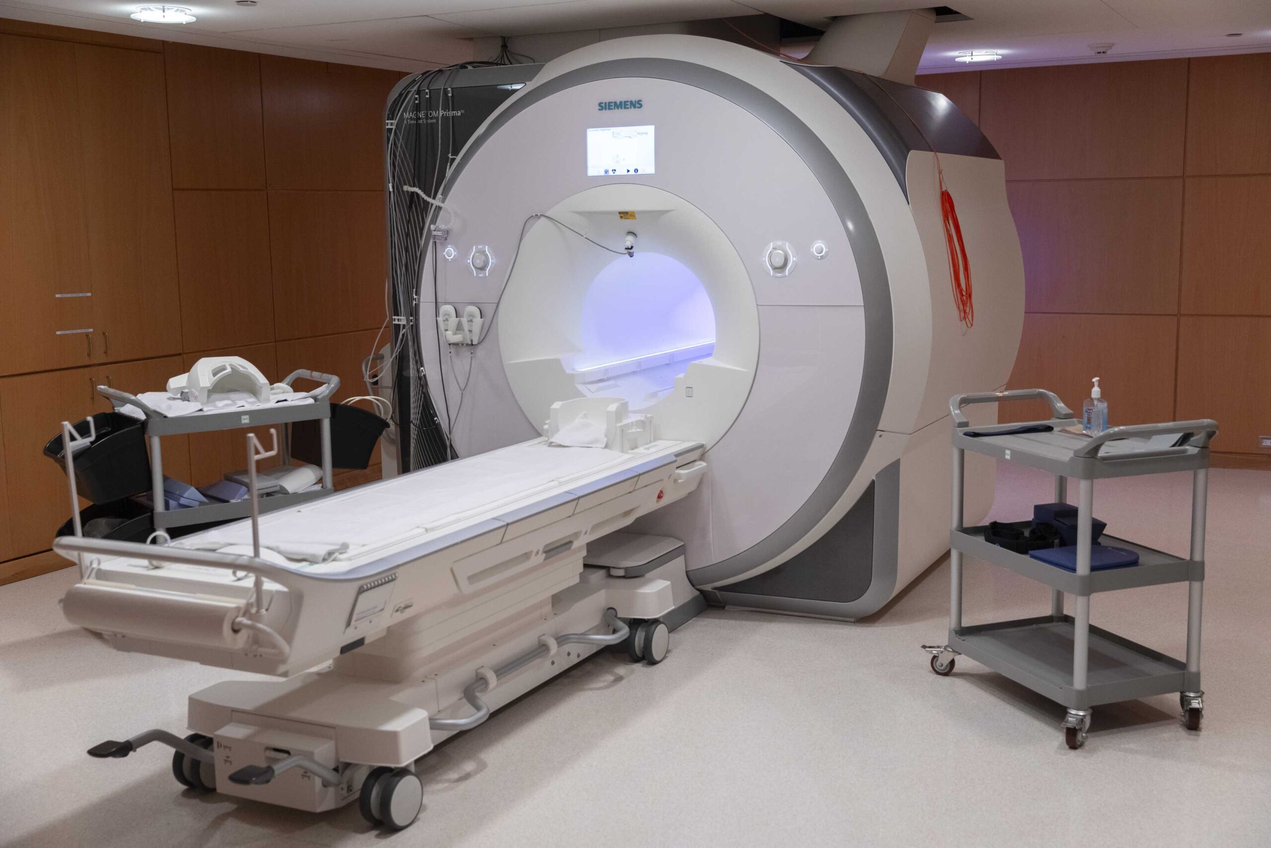
Magnetic resonance imaging (MRI) is a non-invasive imaging technique that uses radio waves and water molecules in the brain to construct brain images. MRI can scan through the skull and show a high level of detail about the brain, without injection or radioactivity.
MRI allows researchers to study multiple aspects of the aging brain. These range from the fibres that connect different parts of the brain, to the blood vessels that sustain it and connect it to other areas of the body. Particularly powerful is MRI’s ability to map brain blood flow and oxygen use for the early detection of dementia.
MRI is used at Baycrest in all aspects of dementia research and is also used to study healthy cognitive functioning, late-life depression, traumatic brain injury, stroke and brain damage resulting from diabetes.
Researchers and scientists at Baycrest @link are pioneering the use of MRI to help evaluate and validate new brain therapies, helping to shorten the time from research findings to practical applications.
Learn more about how Baycrest uses MRI in research:
Magnetoencephalography (MEG) is an advanced neuroimaging technology that measures tiny magnetic fields produced by electrical activity in the brain. Hundreds of sensors arranged in a helmet pick up these magnetic fields, and sophisticated algorithms transform this information into a high-resolution map of neural activity.
Researchers use MEG to study brain activity involved in vision, hearing, memory, motor function and higher cognitive abilities. The signal obtained while participants simply rest is also informative, especially in clinical disorders such as stroke and Alzheimer’s disease.
MEG is especially “participant-friendly” in that it is silent and requires only minimal set-up. Baycrest researchers use MEG to study changes in brain activity linked to dementia — changes that can appear years before symptoms are noticeable. This makes MEG a valuable tool for detecting dementia early and for testing treatments that aim to slow its progression.
Baycrest is one of only six sites in Canada equipped with MEG technology, among about 100 centres worldwide. The in-house combination of MEG with other neuroscience technologies such as MRI, EEG, TMS and eye tracking highlights Baycrest’s unique and advanced research capability.
Learn more about how Baycrest uses MEG in research:
Baycrest study uncovers promising new target for stroke treatment
Baycrest also houses a Hyperfine Swoop system, which obtained Health Canada approval, and is the only portable low-field MRI that is commercially available and clinically approved in the world. This is a 64 mT MRI system with a footprint of 86 cm x 86 cm – similar to an average refrigerator. Moreover, it is mobile, and can be moved by a single operator to the bedside across different spaces, and even transported by specialized vehicles to sites across the region. The low field strength means that electronic devices can operate in the immediate vicinity of this system, and that no shielded room is required. Despite its size and portability, this system can produce T1-weighted MRI, T2-weighted MRI, T2-weighted fluid-attenuated inversion-recovery (T2-FLAIR) and diffusion–weighted MRI (DWI) images. The scan times for a low-field MRI protocol are in fact similar to those of high-field MRI protocols. Thus, we can bring MRI into more communities and reach more diverse study cohorts.
Transcranial direct current stimulation (tDCS) is a non-invasive method of electrical brain stimulation. Specialized electrodes are attached to specific points of the scalp to stimulate brain activity while participants engage in common activities they are trying to improve. For example, to improve motor abilities, electrodes are placed over parts of the brain that control motor function while the participant conducts a motor activity, such as playing a board game or walking.
Scientists at the Rotman Research Institute are investigating tDCS as a potential therapy for people who are impaired in a particular domain, such as language or memory, motor function or gait (walking). As well, it is being used with stroke survivors to help them regain lost abilities and recover faster.
There is also promising research showing that tDCS could be used to treat symptoms of dementia and possibly slow down its progression. Baycrest is conducting studies examining how tDCS could be used to improve memory, general cognition and the ability to name objects in people living with Alzheimer’s disease, primary progressive aphasia, mild cognitive impairments or frontotemporal dementia, as well as motor conditions including Parkinson’s disease and progressive supranuclear palsy.
Some of the ways Baycrest research uses tDCS:
tDCS can produce neurological changes
Participants benefitting from receiving tDCS
Speech therapy combined with tDCS leads to better naming ability than speech therapy only
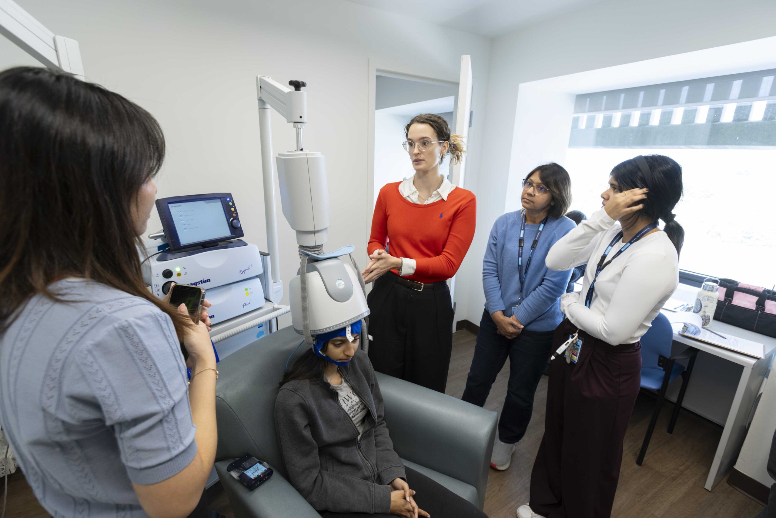
Transcranial magnetic stimulation (TMS) is a non-invasive brain stimulation technique that induces an electrical current in targeted areas of the brain. TMS can be used to study brain function by probing how stimulation influences behaviour when applied to specific brain areas at specific times. The brain’s responses to TMS can be affected by certain neurological disorders, allowing researchers to track disorder severity and assess the impact of treatments.
When repeated over many days, brain stimulation can cause a long-lasting change in brain function and can be used therapeutically to treat disorders. TMS may be used in the treatment of depression and is being explored in the care of movement disorders, stroke, dementia and other conditions. It can also be used with transcranial direct current stimulation (TDCS), a simpler and portable technology with more potential for at-home use.
One of the ways Baycrest research uses TMS:
Baycrest pioneering brain stimulation therapy to target aphasia in recovering stroke patients
Baycrest TMS equipment and facilities
- TMS – Magstim Super Rapid Plus stimulator for rTMS, theta burst and other repetitive paradigms
- Magstim Bistim for paired-pulse and single-pulse experiments
- Brainsight Neuronavigation system
- Brainsway H1-coil deep TMS (dTMS) system – used in current studies of experimental treatment of depression and dementia
- HD-TDCS: Neuroconn 8-channel stimulator, also equipped for TACS, TRNS and custom waveform delivery – MRI and MEG compatible
- TMS-compatible EEG system: Truscan, 128-channel, simultaneous measurement capability
- EMG acquisition system for motor-related studies
Vision is often considered to be the most important sense, and good vision is crucial for maintaining quality of life in older age.
In the Vision Lab, researchers use different methods to study vision and visual perception.
Traditional charts used during eye tests evaluate the ability to see small details and differences in depth, and very faint stimuli.
In addition to traditional charts, Baycrest researchers use sophisticated visual displays such as VIEWPixx – VPixx Technologies to show visual stimuli with great precision and control over factors such as duration and colour. This allows researchers to evaluate visual processes such as the perception of motion, depth, facial identity, emotion and colour. These visual functions can be affected in healthy aging, and some of them can be specifically impacted in individuals with dementia or those with eye diseases such as cataracts, glaucoma or macular degeneration.
The sophisticated displays are also used with other equipment, such as eye tracking devices, electroencephalography (EEG), response buttons and sound equipment, which allow a deeper understanding of visual processing and how it interacts with hearing and memory.
Read more about recent research that made use of the Vision Lab:
The effects of aging on directionally selective masking
The time course of stimulus-specific perceptual learning